The GLP aggregated and excerpted this blog/article to reflect the diversity of news, opinion and analysis.
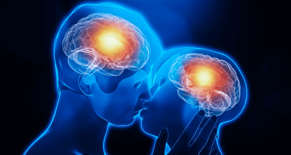
A mother and her child are curled up together inside the tube of a 3 Tesla magnetic resonance imaging scanner in April 2015. The scanner bangs and beeps, shudders and screeches. The baby is finally sleeping, pressed firmly against his mother’s chest, and so is still enough for the MRI to see inside his head. A single MR image, like this one, takes several minutes to capture. Moving just a millimeter leaves a blur on the screen. The mother and baby must hold their pose, as if for a daguerreotype.
This particular MR image was not made for diagnostic purposes, nor even really for science. No one, to my knowledge, had ever made an MR image of a mother and child. We made this one because we wanted to see it.
Here is a depiction of one of the hardest problems in neuroscience: How will changes in the brain, that specific little organ, accomplish the unfolding of a whole human mind?
Read full, original post: Why I Captured This MRI of a Mother and Child