Doctors used to perform autopsies on nearly half of all patients who died in the hospital in order to understand their cause of death. The number has fallen to just 5 percent of patients in recent years. Some say the shortage of trained pathologists and medical examiners are to blame, while other’s point to the public’s general squeamishness about dissecting their loved ones.
But, forensic pathologists have started using a new, non-invasive technology to determine cause of death and to investigate what happens to the brain after stroke and heart attack. They’re putting corpses in body scanners.
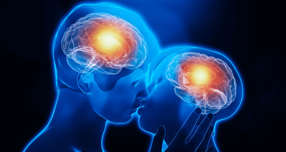
Although they’ve long been used as an important technology for diagnosing the living, computerized tomography (CT) and magnetic resonance imaging (MRI) scanners can be used to identify injury and investigate death from the inside of a patient out. Scanners take a series two-dimensional pictures, called slices, of a patient’s body, then recompile them into a full body image of all the tissues, giving literally the whole picture of what’s recently happened inside. While autopsies offer the chance to take tissue samples and look at death at the microscopic level, whole body scans show the whole picture. And no where is that point of view more important that in the delicate, hard to study and easy to injure brain:
Physician Manuela Baglivo at the Institute of Forensic Medicine in Zurich recently… described how the pervasiveness of fatal head injuries has led to a surge in studies involving brain scanning corpses over the past decade. They also note an increasing interest from other areas of medicine where post-mortem brain scanning is helping to understand the lethal effects of brain injury, such as strokes and internal bleeds, as a way of ultimately helping to save lives.
Studying the brains of dead people also offers scientists the opportunity to test the limits and the accuracy of certain brain scanning technologies, specifically functional Magnetic Resonance Imaging (fMRI). These scans match anatomical data on top of images that follow the signal of blood flow in the brain, which scientists take as a proxy of brain activity. This is how we know what specialized brain areas are activated when we look at human faces compared to other objects, or which areas increase in activity when we are angry or upset.
fMRI is considered a vital tool for the large-scale brain initiatives currently underway in the U.S. and U.K. to help map the brain and its connections. But the technology involves a lot of estimation, extrapolation and mathematical correction, so its sometimes difficult to tell if you’re getting a true signal brain activity or an artifact. Studying corpses, who definitively have no brain activity, is a way to test this. Vaughan Bell at the Guardian recently described a study to test if a type of electrical brain stimulation was causing false positives in fMRI studies: “They found that if they applied the stimulation to their dead subjects they seemed to show brain activity where there was none, providing an important caution for those researching the technique on the living.”
Meredith Knight is a blogger for Genetic Literacy Project and a freelance science and health writer in Austin, Texas. Follow her @meremereknight.
Additional Resources:
- MIT researchers use MRI to study genetic activity in brains, Genetic Literacy Project
- Risk genes show up in newborns’ brain scans, Futurity